나이를 기준으로 시술 계획을 짜도 될까요. 저는 상담에서 나이 숫자보다 지금 앞에 있는 얼굴을 먼저 봅니다. 나이는 물어봐야 알 수 있지만, 얼굴은 묻지 않아도 바로 보이기 때문입니다.
피부, 지방, 근육, 뼈가 함께 변한다는 건 Fitzgerald 등의 「Update on facial aging」(Aesthet Surg J, 2010)에도 정리돼 있습니다. 제 진료에서는 그 변화의 속도와 양상이 사람마다 달랐습니다. 동갑이어도 얼굴이 늙는 모양은 제각각이에요.
앞뺨이 처져 보이면 조직이 그만큼 내려온 걸까요? Rohrich 등(Plast Reconstr Surg, 2008)은 심부 내측 뺨 지방이 줄면 앞뺨이 실제보다 처져 보이는 가성 하수가 생기고, 팔자주름도 더 도드라져 보일 수 있다고 해석했습니다. Lambros(Plast Reconstr Surg, 2007)는 같은 사람들의 사진을 10~50년 간격으로 비교했습니다. 눈꺼풀과 뺨의 경계 같은 중안부 위쪽 기준점의 높이는 비교적 그대로였습니다.
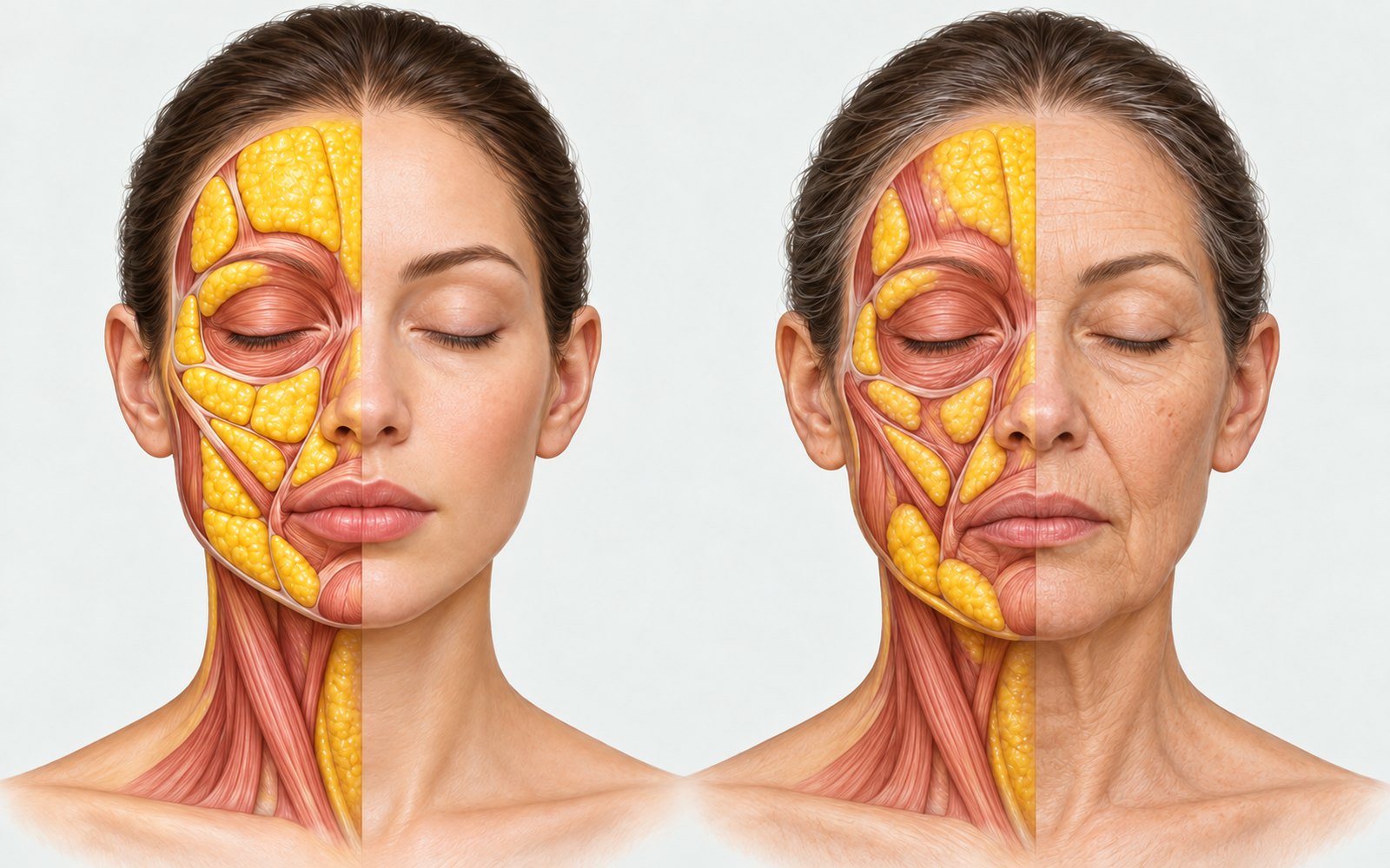
지방 구획의 변화(설명용 일러스트). 나이가 들면 지방 구획의 볼륨이 줄고 아래로 옮겨 갑니다. 왼쪽이 젊을 때, 오른쪽이 노화 후입니다.
그래서 처져 보이는 얼굴이라도 볼륨이 빠지고 옮겨 간 건 아닌지 같이 봅니다. 볼륨이 줄었거나 위치가 달라진 부위를 먼저 확인하고, 무엇을 더할지는 그다음에 정합니다.
토대인 뼈부터 보면, 상악골과 이상구 주변이 흡수되고 하악도 높이와 길이가 줄어요. 그 위의 연부조직이 그대로여도 얼굴이 꺼지고 처져 보이는 까닭입니다. 지방 구획은 줄거나 아래로 내려앉고, 피부에서는 진피 콜라겐이 줄어 결은 거칠어지고 되돌아오는 힘은 약해집니다. 근거 문헌은 백서 3장 본문에 번호로 달고 끝의 참고문헌에 모아 두었습니다.
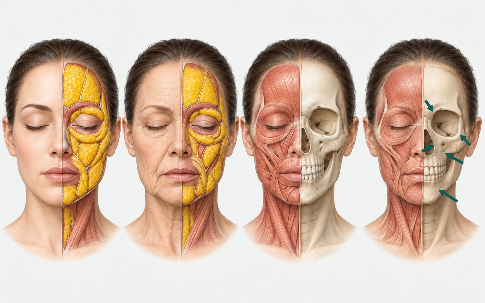
노화에 따른 얼굴 층의 변화(설명용 일러스트). 왼쪽 두 그림은 피부와 지방, 오른쪽 두 그림은 근육과 뼈이며 각각 젊을 때와 노화 후입니다. 오른쪽 끝 그림의 화살표가 뼈 흡수가 일어나는 부위입니다.
제가 본 얼굴들에서는 유지 인대가 고정한 곳이 비교적 덜 움직이고, 인대와 인대 사이가 처져 보이는 경우가 많았습니다. 턱선의 jowl이 그 예죠.
볼륨이 빠진 곳에는 그림자가 집니다. 저는 같은 조명에서 정면과 좌우 측면, 좌우 45도로 찍고, 턱을 든 각도와 위에서 내려다본 각도도 따로 찍습니다. 정면에서 놓친 꺼짐과 윤곽의 굴곡이 이 각도들에서 잡힙니다. 자리와 조명이 매번 같아야 다음 회차와 비교할 수 있습니다.
촉진은 헤어라인과 볼, 턱선 피부를 살짝 집어 당겼다 놓는 식으로 하고, 복원이 더딘 곳을 탄력 저하로 봅니다. 다만 손으로 알 수 있는 건 주로 탄력과 이완이에요. 볼륨이 빠졌는지 판단하려면 사진의 그림자도 함께 봐야 합니다. 시술 이력과 병력도 사진, 촉진과 같은 비중으로 봅니다.
빠진 곳을 찾았다고 한 회에 몰아서 채우면 얼굴이 무거워 보이기 쉬워요. 저는 여러 회차에 나눠 쌓고, 회차마다 같은 조건의 사진으로 확인합니다.
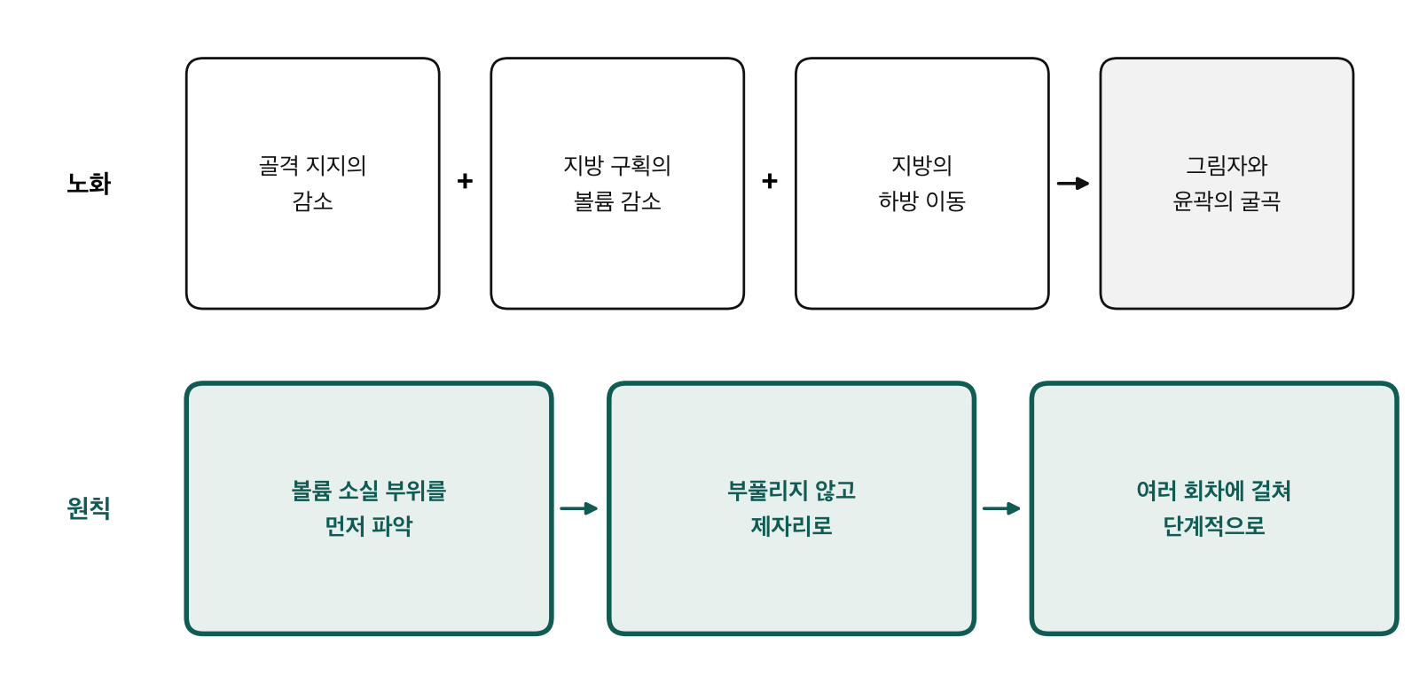
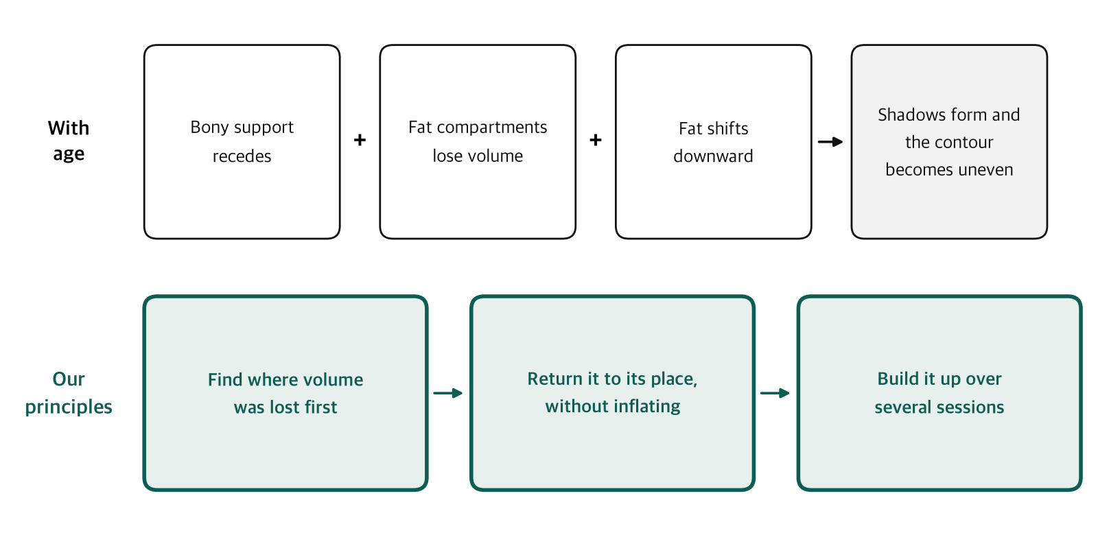
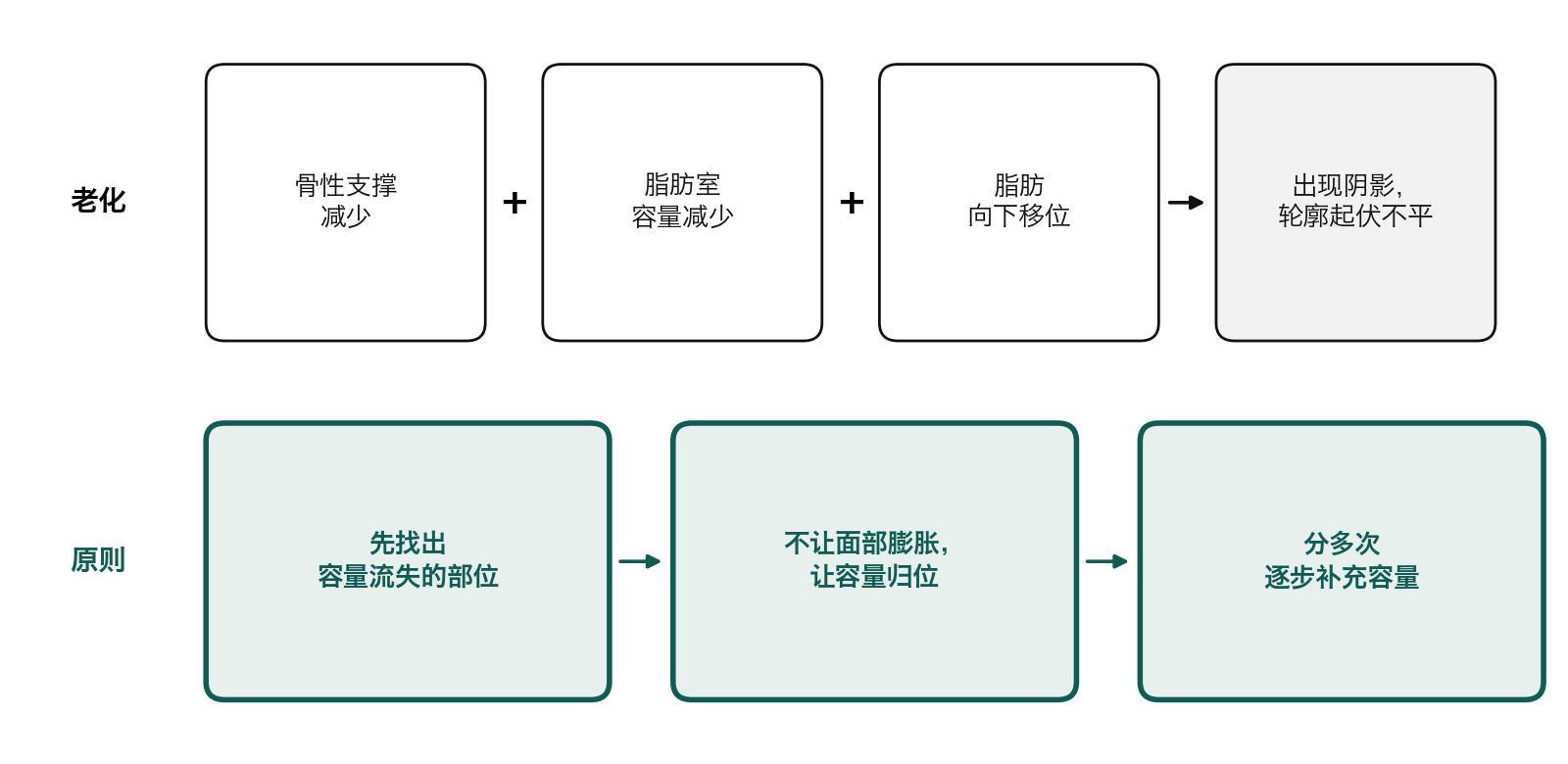
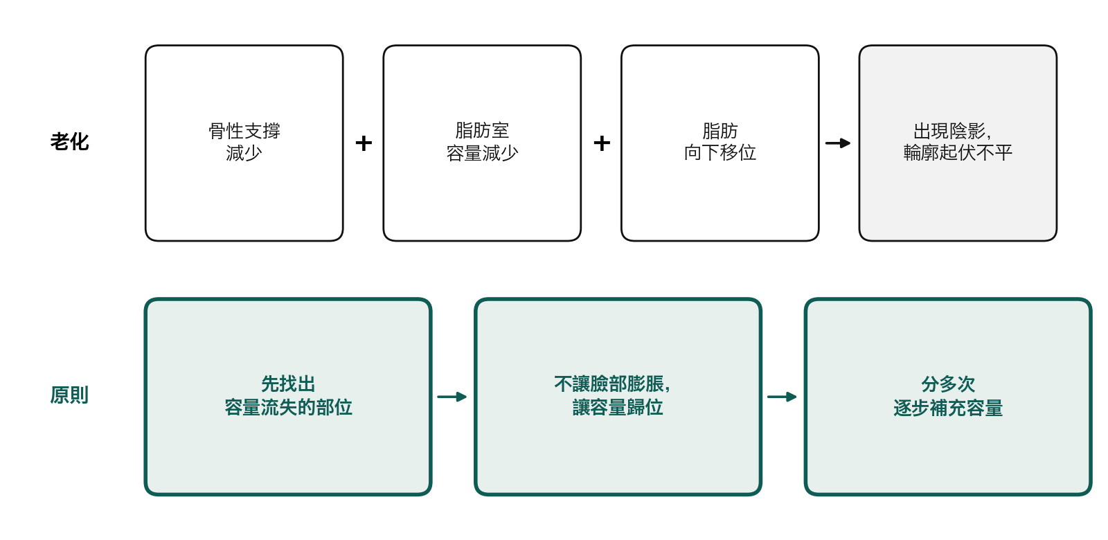
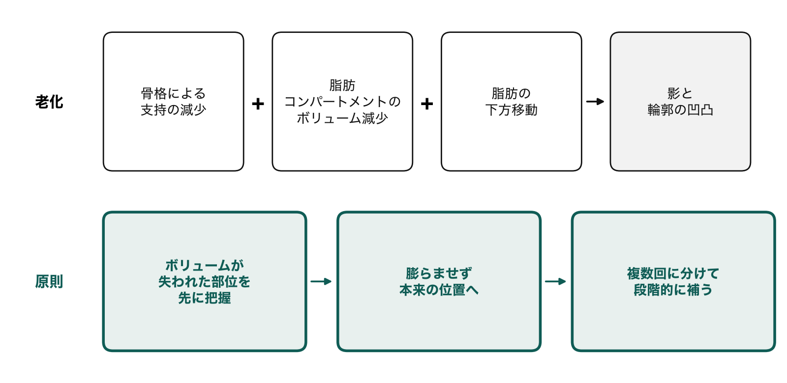
노화에 따른 얼굴의 변화(위)와 볼륨 복원의 원칙(아래).
시술 당일 피부 컨디션이 어떤지, 며칠 안에 어떤 일정이 있는지 먼저 확인하고, 애매하면 미룹니다. 어떤 경우에 시술하고 언제 미룰지는 교육 때 구체적으로 다룹니다.
Can you plan a treatment around a patient's age? In consultations I look at the face in front of me before the number. I have to ask a patient's age, but I can see the face right away.
That skin, fat, muscle, and bone all change together is well summarized in Fitzgerald et al., "Update on facial aging" (Aesthet Surg J, 2010). In my practice, the pace and pattern of that change have differed from person to person. Two people the same age can age in very different ways.
When the anterior cheek looks lower, has the tissue really descended that far? Rohrich et al. (Plast Reconstr Surg, 2008) suggested that loss of the deep medial cheek fat can create pseudoptosis, making the anterior cheek look lower than it is and the nasolabial fold more prominent. Lambros (Plast Reconstr Surg, 2007) compared photographs of the same people taken 10 to 50 years apart. Landmarks in the upper midface, such as the lid–cheek junction, stayed at roughly the same height.
Fat compartments with age (illustration): they lose volume and shift downward. Left, young; right, aged.
So even when a face looks like it has dropped, I check whether volume has been lost or has shifted. I first identify where volume has decreased or moved, and only then decide what to add.
Start with the foundation. The maxilla and the area around the piriform aperture resorb, and the mandible loses height and length. That is why a face can look hollow and saggy even when the soft tissue over it has not changed. Fat compartments shrink or settle downward, and in the skin, dermal collagen decreases, so texture coarsens and recoil weakens. The supporting references are numbered in chapter 3 of the white paper and collected in its reference list.
How the facial layers change with age (illustration). Left pair: skin and fat; right pair: muscle and bone; each shown young, then aged. Arrows in the far-right image mark sites of bone resorption.
In the faces I have seen, areas held by the retaining ligaments tend to move less, while the spaces between ligaments often look as if they have dropped. The jowl along the jawline is one example.
Where volume is lost, a shadow forms. Under the same lighting, I take frontal, right and left profile, and right and left 45-degree photographs, plus separate views with the chin raised and from above. These views catch hollows and contour irregularities the frontal view misses. The position and lighting have to be the same every time so you can compare against the next session.
For palpation, I lightly pinch the skin at the hairline, cheek, and jawline, pull, and release, and I interpret slow recoil as a sign of reduced elasticity. But what your hands can tell you is mostly elasticity and laxity. To judge whether volume has been lost, you also need the shadows in the photographs. I give treatment history and medical history the same weight as photographs and palpation.
Even after identifying areas of volume loss, filling them all in a single session can easily make the face look heavy. I restore volume gradually over several sessions, assessing the face at each session with photographs taken under the same conditions.
Top: how the face changes with age. Bottom: the principles of restoring volume.
On the day of treatment, I first check the condition of the skin and whether the patient has anything scheduled in the next few days. If I am unsure whether to proceed, I postpone treatment. During training, I discuss in detail when to treat and when to postpone.
This article expands on chapter 1 of the white paper "A Face Looks Smaller When Volume Is in Its Place." The full white paper and its references are at www.vpmedi.com/whitepaper/.