A Face Looks Smaller When Volume Is in Its Place
Principles of facial volume in aging — a clinical perspective from more than a decade of aesthetic practice
Seong-Pil Jang, M.D. · Medical Director, Ellis Clinic (since 2014) · CEO, Veripuremedi Co., Ltd. — a physician who loves electronic engineering and keeps researching it
Summary
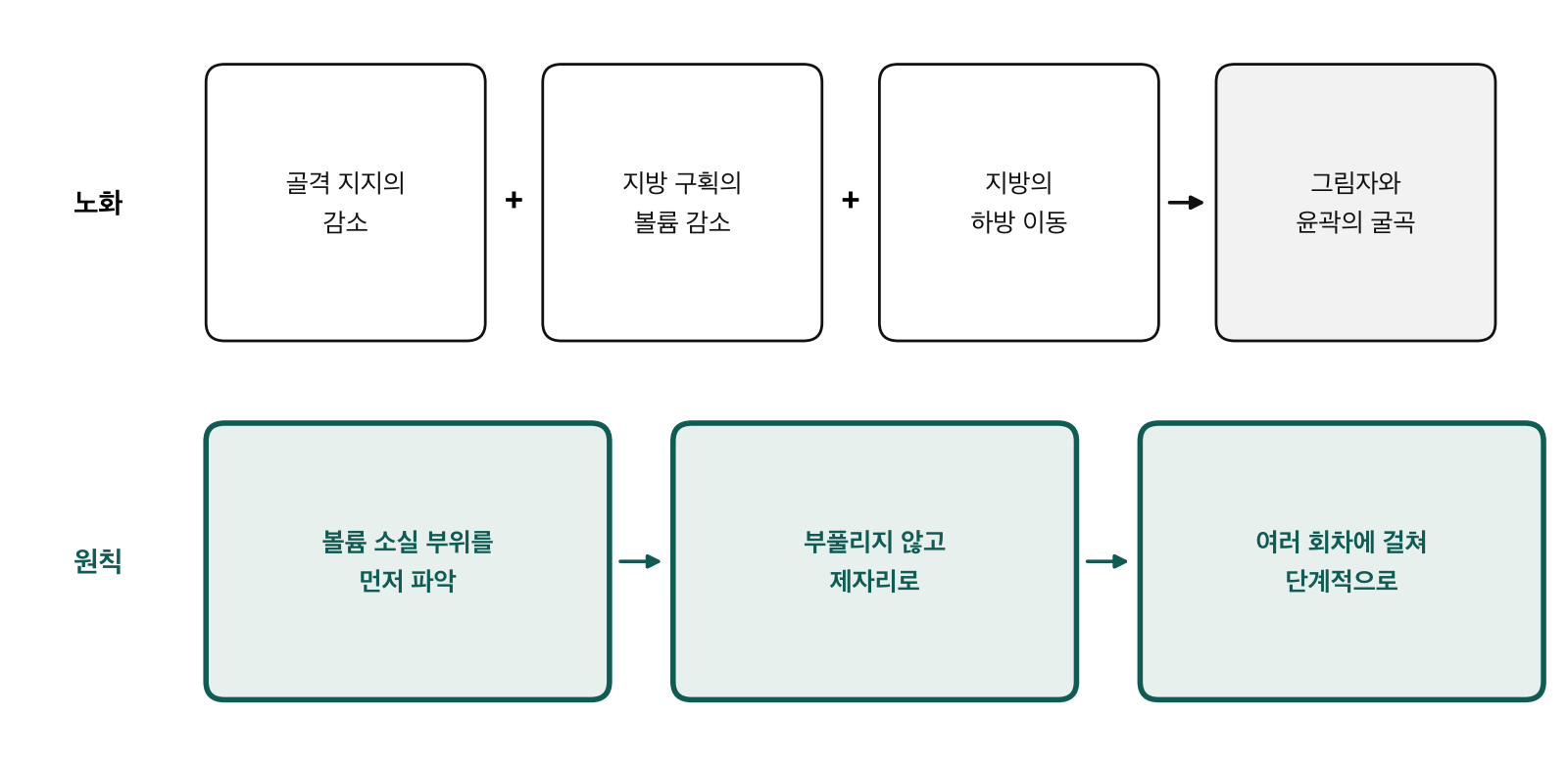
Bone size alone does not determine how small a face looks. Where the volume sits matters a great deal. With age, skin, fat, muscle, and bone all change together. Bony support recedes, fat compartments lose or shift volume, and dermal collagen declines, so the outline of the cheekbones and jaw begins to show. In our practice, these faces look longer, more angular, and even larger.
So the principle we follow is simple. We do not inflate the face. We first identify where volume has been lost, return volume to those areas, and build it gradually over several sessions. This paper explains the anatomy behind that principle and how we assess a face.
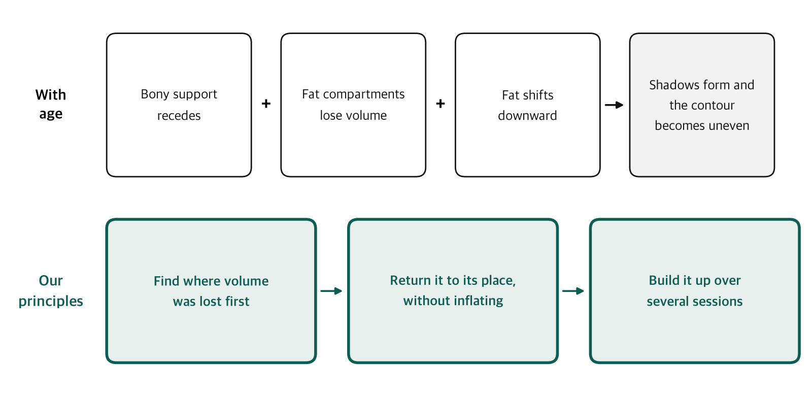
Figure 1. Top: how the face changes with age. Bottom: the principles we follow in restoring volume.
1. Aging Arrives in Stages
Aging does not arrive all at once. Age-related changes involve skin, fat, muscle, and bone [8]. In our practice, their pace and pattern vary from person to person. That is why two people of the same age can age very differently, and why we look at the face, not the age. We start by looking at what has been lost and what has shifted in this person's face.
2. The Triangle of Youth Inverts
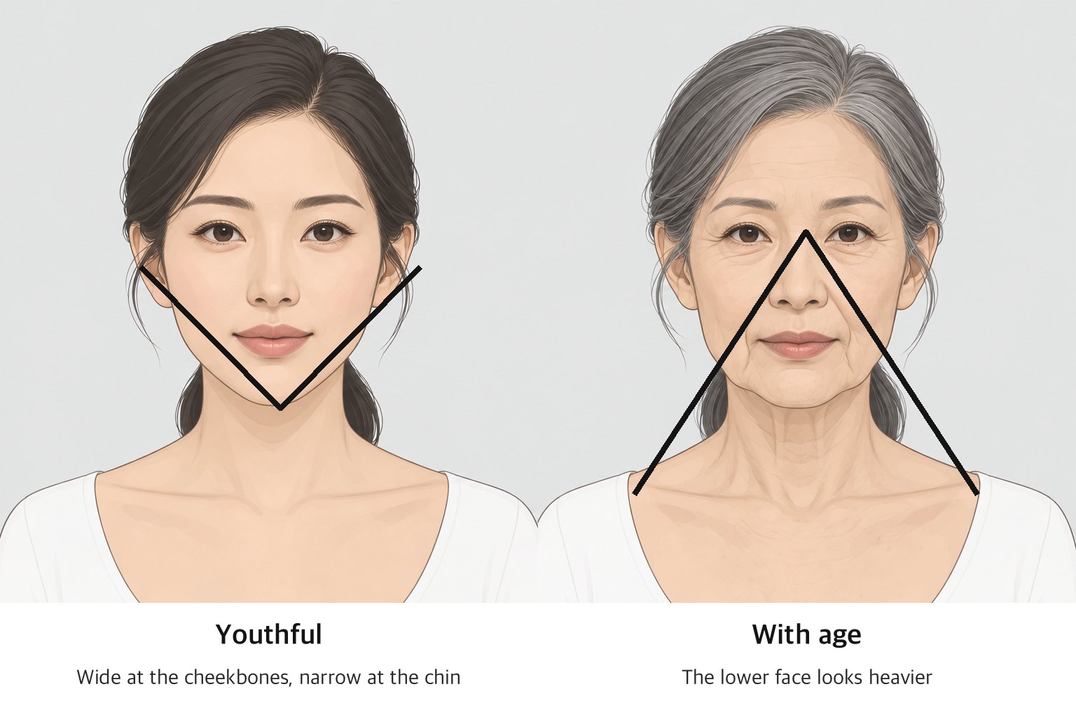
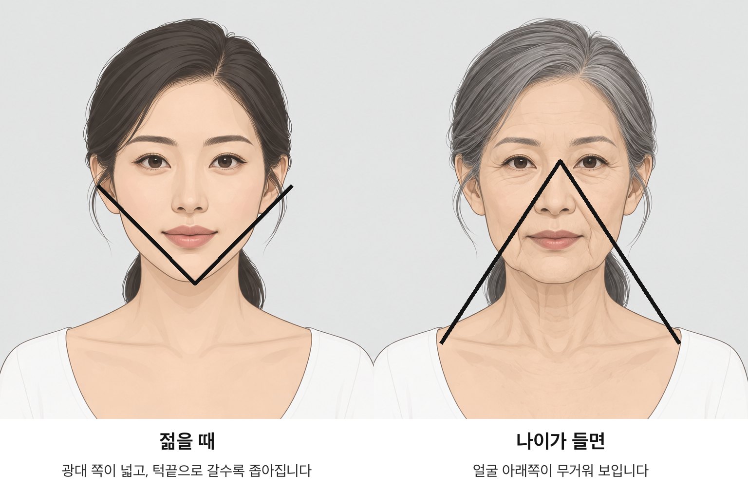
Figure 2. The triangle of youth: wide at the cheekbones when young; with age, the wide end of the triangle moves down.
The "triangle of youth," long used in aesthetic medicine, captures this change at a glance. A young face is wide at the cheekbones and narrows toward the chin, an inverted triangle. As the anterior cheek loses volume and weight settles lower, the wide end of the triangle moves down. The jawline blurs and the lower face looks heavy. We aim for a balanced distribution of facial volume, with the wide end of the triangle back at the top.
3. Every Layer Changes Together
| Layer | With age | What it looks like |
|---|---|---|
| Bone | The maxilla, the piriform aperture region, and parts of the orbital rim resorb; the mandible loses height and length [4][5][9] | Weaker support at the anterior cheek and beside the nose; a softer jawline |
| Fat | Fat compartments lose volume or shift downward [1][3] | Anterior cheek hollows; the nasolabial fold can look more prominent [2] |
| Ligaments | Retaining ligaments separate spaces and compartments [6]; in our practice, tissue between them appears to descend | Jowling and contour breaks along the jawline |
| Skin | Dermal collagen declines with age [10] | Rougher texture, less recoil |
3.1 Bone: The Foundation Shrinks
Facial bone does not shrink evenly with age; it recedes differently from region to region. The maxilla and the bone beside the piriform aperture resorb and recede, and so do the upper inner and lower outer parts of the orbital rim [4][5]. The mandible also loses height and length, weakening the support behind the jawline [9]. As the bony foundation beneath the skin and fat shrinks, the face looks more hollow and saggy, even when the soft tissue above stays the same.
3.2 Fat: It Shrinks and Moves
Facial adipose tissue is organized into distinct anatomical compartments separated by septa [1]. Cadaver CT comparisons have shown downward displacement of fat compartments, an inferior volume shift within compartments, and volume loss in selected compartments with age [3]. The authors of a related study link loss of the deep medial cheek fat to pseudoptosis of the cheek and a more prominent nasolabial fold [2].
Photographic comparisons of the same individuals over 10 to 50 years found relative vertical stability of the lid–cheek junction and other upper-midface landmarks [7]. Based on findings like these, we see aging not only as sagging but as volume being lost and moving out of place.
3.3 Ligaments and the Jawline: Where Tissue Is Held and Where It Falls
The retaining ligaments of the face separate its anatomical spaces and compartments [6]. In our practice, tissue held by a ligament tends to stay put, while tissue between ligaments is more likely to appear to descend. Jowling along the jawline is an example. The mandibular septum separates jowl fat from submandibular fat [11]. Changes on either side of that boundary, together with loss of skin elasticity, break the smooth line of the jaw.
3.4 Skin: Collagen Declines, and Thickness Varies by Area
Dermal collagen declines with age. Measurements of forearm skin showed an age-associated decline in collagen content [10], and reduced collagen synthesis in aged skin involves both changes in fibroblast function and reduced mechanical stimulation [12]. We see these changes as underlying skin that thins and loses its recoil.
Facial skin thickness also varies by area. In a cadaver study, total skin thickness was lowest at the upper medial eyelid and highest at the lower nasal sidewall [13]. The same principle has to be applied differently from one area to the next.
4. Why a Hollow Face Can Look Larger
This section reflects what we have observed over more than a decade of practice. When volume is gone, the outline of the cheekbones and jaw shows through. An uneven contour makes the face look angular, and hollowing at the front of the cheeks makes the midface look longer. So a face that has lost volume can look not only older but larger. In faces that look young and small, we consistently see a short midface and a smooth, continuous outline. In our practice, this has held across ethnicities.
5. The Principle: Return, Do Not Inflate
Start with the anterior cheek. When we assess midfacial proportions, we look first at anterior cheek fullness. What we aim for is a visually shorter midface.
Assess the outline of the face. The frame that runs from the hairline around the ear and along the jawline is the first thing to soften when elasticity drops. We assess temporal and lateral cheek hollowing together with that outline. The outline we aim for is a smooth, continuous contour with a gently tapered lower face, a soft V-line.
Know where to stop. Fill the forehead without making it bulge. Keep the lips full but not overdone. Leave the nose alone, and keep the natural shape of the eyes.
Build in stages. We prefer to build volume over several sessions rather than all at once, because inflating a face in one sitting tends to make it look heavy. Hyaluronic acid is highly hygroscopic and is degraded in the body by hyaluronidases and oxidative stress [14]. So we distinguish the initial volume from the changes in the tissue that follow, and we track them separately.
6. How to Identify Areas of Volume Loss
Assess volume in photographs. Lost volume shows up as shadow. Photographs taken under the same lighting, from the front, from each side, at 45 degrees on each side, with the chin raised, and from above, show hollows and uneven contours that a single front-facing photo misses. We take photographs in the same spot under the same light each time, so each session can be compared with the last.
Assess by palpation. Gently pinch and pull the skin at the hairline, cheek, and jawline. Where it is slow to spring back, elasticity has been lost. Palpation tells us about laxity. We identify areas of volume loss by weighing what we feel together with the shadows in the photographs.
Review the history. We give the patient's treatment and medical history the same weight. We start by identifying where volume is missing before deciding what to add.
7. The Person Before the Procedure
We look at the person before the procedure. We ask first about current skin condition, medical history, past treatments, and even the next few days' schedule. When in doubt, we do not treat that day. Specific criteria for who to treat and when to wait are covered in our education program.
8. Working with Devices: Stimulating Tissue and Restoring Volume
Energy-based devices such as HIFU and monopolar RF tighten tissue with heat and stimulate collagen production. Volume restoration fills in the volume that is missing. One stimulates tissue and the other restores volume, so neither replaces the other. We see volume restoration as an addition to a clinic's existing use of energy-based devices.
9. From the Clinic to the Home
Treatments are occasional; home care is daily. We think of in-clinic assessment and daily skin care as one continuous process. That is why we develop a clinic-only line and a home-care line together.
About This Paper
This paper discusses the principles of facial aging and volume distribution only. Clinical technique is taught directly in our invitation-only education program.
About Veripuremedi
Veripuremedi is a skincare and medical-device company born in aesthetic practice. We make LIZ VERIPURE PRO, a clinic-only line (eight solutions: five available now, three in development), and LIZ VERIPURE CARE, a seven-product home-care line, and we file our own patents on low-pain medical devices.
Contact: care@vpmedi.com · www.vpmedi.com · liz.vpmedi.com · Instagram @veripuremedi
Veripuremedi Co., Ltd. · Ulsan, Republic of Korea
References
- Rohrich RJ, Pessa JE. The fat compartments of the face: anatomy and clinical implications for cosmetic surgery. Plast Reconstr Surg. 2007;119(7):2219–2227.
- Rohrich RJ, Pessa JE, Ristow B. The youthful cheek and the deep medial fat compartment. Plast Reconstr Surg. 2008;121(6):2107–2112.
- Gierloff M, Stöhring C, Buder T, Gassling V, Açil Y, Wiltfang J. Aging changes of the midfacial fat compartments: a computed tomographic study. Plast Reconstr Surg. 2012;129(1):263–273.
- Shaw RB Jr, Kahn DM. Aging of the midface bony elements: a three-dimensional computed tomographic study. Plast Reconstr Surg. 2007;119(2):675–681.
- Mendelson B, Wong CH. Changes in the facial skeleton with aging: implications and clinical applications in facial rejuvenation. Aesthetic Plast Surg. 2012;36(4):753–760.
- Alghoul M, Codner MA. Retaining ligaments of the face: review of anatomy and clinical applications. Aesthet Surg J. 2013;33(6):769–782.
- Lambros V. Observations on periorbital and midface aging. Plast Reconstr Surg. 2007;120(5):1367–1376.
- Fitzgerald R, Graivier MH, Kane M, et al. Update on facial aging. Aesthet Surg J. 2010;30(Suppl):11S–24S.
- Shaw RB Jr, Katzel EB, Koltz PF, et al. Aging of the mandible and its aesthetic implications. Plast Reconstr Surg. 2010;125(1):332–342.
- Shuster S, Black MM, McVitie E. The influence of age and sex on skin thickness, skin collagen and density. Br J Dermatol. 1975;93(6):639–643.
- Reece EM, Pessa JE, Rohrich RJ. The mandibular septum: anatomical observations of the jowls in aging — implications for facial rejuvenation. Plast Reconstr Surg. 2008;121(4):1414–1420.
- Varani J, Dame MK, Rittie L, et al. Decreased collagen production in chronologically aged skin: roles of age-dependent alteration in fibroblast function and defective mechanical stimulation. Am J Pathol. 2006;168(6):1861–1868.
- Chopra K, Calva D, Sosin M, et al. A comprehensive examination of topographic thickness of skin in the human face. Aesthet Surg J. 2015;35(8):1007–1013.
- Fallacara A, Baldini E, Manfredini S, Vertuani S. Hyaluronic acid in the third millennium. Polymers (Basel). 2018;10(7):701.
Update history
- Updated the author line.
- First published.